Acridine Orange is a versatile metachromatic dye that interacts differently with various cellular components. When used at low concentrations, it intercalates with DNA while causing RNA precipitation. At higher concentrations, however, it denatures and precipitates both RNA and DNA.
This compound is particularly valuable for autophagy analysis due to its pH-sensitive properties. Acridine Orange enters acidic organelles in a pH-dependent manner, producing distinct fluorescence patterns: green fluorescence at neutral pH and bright red fluorescence in acidic conditions as it accumulates within acidic organelles.
Beyond nucleic acids, Acridine Orange can also stain acid glycosaminoglycans, making it a multifunctional tool in cellular analysis.
Kundu S, Kim TH, Yoon JH, Shin HS, Lee J, Jung JH, Kim HS. Viriditoxin regulates apoptosis and autophagy via mitotic catastrophe and microtubule formation in human prostate cancer cells. Int J Oncol. 2014 Dec;45(6):2331-40. doi: 10.3892/ijo.2014.2659. Epub 2014 Sep 17. PMID: 25231051.
Vucicevic L, Misirkic-Marjanovic M, Paunovic V, Kravic-Stevovic T, Martinovic T, Ciric D, Maric N, Petricevic S, Harhaji-Trajkovic L, Bumbasirevic V, Trajkovic V. Autophagy inhibition uncovers the neurotoxic action of the antipsychotic drug olanzapine. Autophagy. 2014;10(12):2362-78. doi: 10.4161/15548627.2014.984270. PMID: 25551567; PMCID: PMC4502661.
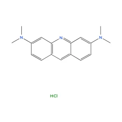
Acridine Orange: The Versatile Fluorescent Dye in Scientific Research
Acridine Orange is a remarkable and versatile fluorescent dye that has become an essential tool in scientific research. Its unique ability to stain DNA, RNA, and acidic organelles makes it invaluable in various biological and medical applications.
One of the most fascinating aspects of Acridine Orange is its pH-dependent fluorescence. In neutral environments, it emits a bright green glow, but in acidic conditions, such as inside lysosomes and autophagic vesicles, it shifts to an intense red fluorescence. This property has made it a key dye for studying autophagy, a cellular process essential for maintaining cell health and combating diseases like cancer and neurodegeneration (Thomé et al., 2016).
Beyond autophagy research, Acridine Orange has proven useful in cancer imaging and therapy. Studies have shown that it can selectively accumulate in cancerous tissues, providing a potential method for enhancing tumor visualization during surgery (Byvaltsev et al., 2019). This makes it a promising tool for improving surgical precision and patient outcomes.
In addition to its medical applications, Acridine Orange has played a crucial role in environmental science. Researchers have investigated its interaction with materials like graphene oxide to develop efficient methods for removing toxic dyes from wastewater (Sun & Fugetsu, 2013). These studies highlight its potential in pollution control and sustainable chemistry.
Acridine Orange is also widely used in cell viability assays and microscopy, helping scientists distinguish between live and dead cells. When used alongside propidium iodide, it allows for precise fluorescence-based measurements of cell health, which is crucial in toxicology and drug testing (Miller et al., 2019).
Furthermore, its interaction with DNA has led to important discoveries in molecular biology and nanotechnology. Recent research has explored how Acridine Orange derivatives can bind to and stabilize G-quadruplex structures in genes like KRAS, which are implicated in cancer development (Bregadze et al., 2014). These findings open up new possibilities for targeted cancer therapies.
From basic cellular biology to advanced medical imaging and environmental protection, Acridine Orange continues to be a powerful tool in scientific exploration. Its versatility and unique fluorescence properties ensure that it will remain a cornerstone of biological and chemical research for years to come.
-
Byvaltsev, V. A., Bardonova, L. A., Onaka, N. R., Polkin, R. A., Ochkal, S. V., Shepelev, V. V., Aliyev, M. A., & Potapov, A. A. (2019). Acridine Orange: A Review of Novel Applications for Surgical Cancer Imaging and Therapy. Frontiers in Oncology, 9, 925.
-
Thomé, M. P., Filippi-Chiela, E. C., Villodre, E. S., Migliavaca, C. B., Onzi, G. R., Felipe, K. B., & Lenz, G. (2016). Ratiometric analysis of Acridine Orange staining in the study of acidic organelles and autophagy. Journal of Cell Science, 129(24), 4622–4632.
-
Sarnat, H. B. (1985). [Acridine orange: a fluorochrome of nucleic acids for the study of muscle and nerve cells]. Revue Neurologique (Paris), 141(2), 120–127.
-
Miller, H. L., Contera, S., Wollman, A. J. M., Hirst, A., Dunn, K. E., Schroeter, S., O'Connell, D., & Leake, M. C. (2019). Biophysical characterization of DNA origami nanostructures reveals inaccessibility to intercalation binding sites. arXiv preprint arXiv:1911.07022.
-
Bregadze, V. G., Melikishvili, Z. G., Giorgadze, T. G., Jaliashvili, Z. V., Chkhaberidze, J. G., Monaselidze, J. R., & Khuskivadze, T. B. (2013). Forster Resonance Energy Transfer and Laser Fluorescent Analysis of Defects in DNA Double Helix. arXiv preprint arXiv:1306.1846.
-
Bregadze, V. G., Melikishvili, Z. G., & Giorgadze, T. G. (2014). Nanophotonics and DNA: New approaches. arXiv preprint arXiv:1406.7272.
-
Sun, L., & Fugetsu, B. (2013). Effect of encapsulated graphene oxide on alginate-based bead adsorption to remove acridine orange from aqueous solutions. arXiv preprint arXiv:1307.0223.